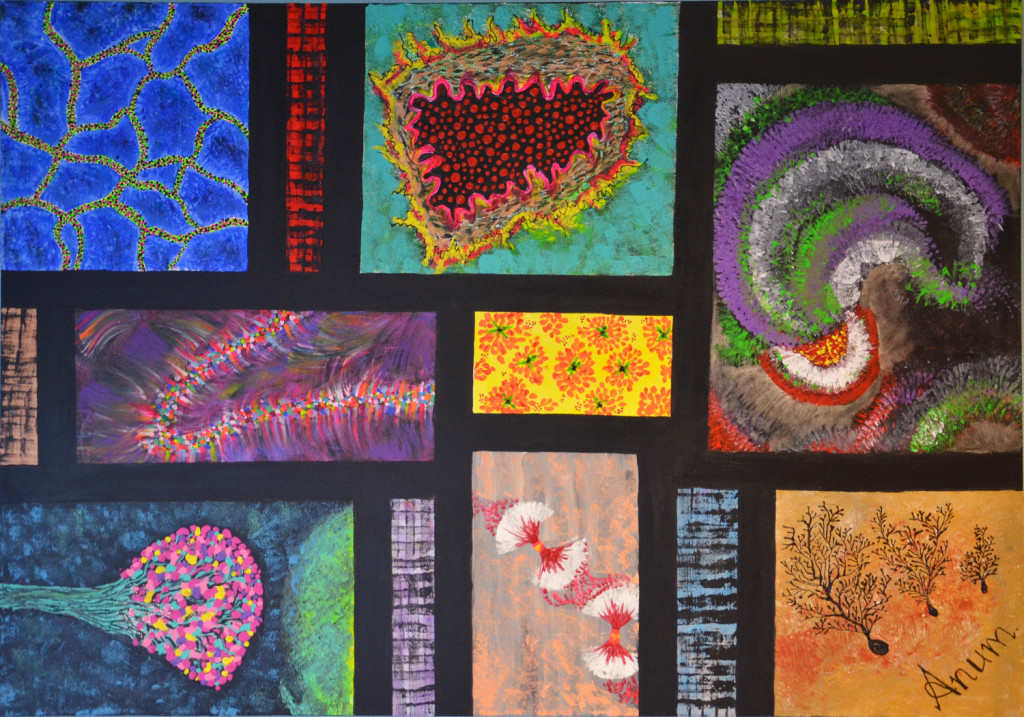
It is a constellation of diverse neuronal images living in color. As an independent artist and medical doctor I am fascinated by artistic expression of scientific excellence. This painting is 100x70x4 cm acrylic painted canvas showcasing in individual sections various aspects of neuroscience based art. On the top left corner the vascular channels in cerebral cortex (blue) are painted. Below this image a brainbow depicting individual neurons of the dentate gyrus projecting their dendrites to the outer layer, where they receive input from the cortex. Below this image a neuronal junction with a presynaptic neuron filled with neurotransmitter vesicles can be visualized. The image in the upper right corner shows the hippocampus of the mouse brain. In the lower right corner cerebellar purkinje cells are drawn with the intricately divided dendrites. The top most image in the center is a cross-section of a brain arteriole filled with red blood cells. The central image in yellow is portraying human neural stem cells forming polarized cellular rosettes with mitotic stem cells at the center. Below this two brain tumor cells are captured in telophase, the final stage of mitosis.
The Neuro Bureau
neuro-collaboration in action

