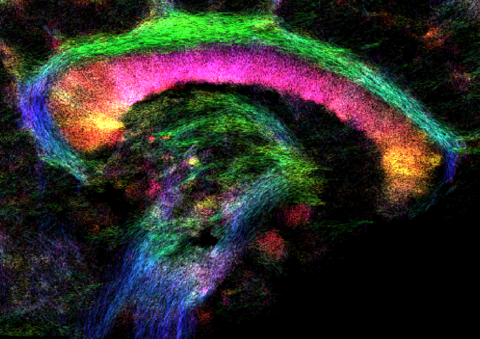
Diffusion MRI using ultra-high field MRI (7 Tesla) depicts parallel white matter fiber structures in vivo at an isotropic spatial resolution of 1mm and below. The image shows a sagittal cut through the cingulum (green), the corpus callosum (red), the thalamus and the brainstem. The texture and the color were generated from the orientation of 3D pathways in the brain. The color corresponds to the orientation of the tracks (red: left-right; green: front-back; blue: top-bottom).