Gabriel Mangeat
NeuroPoly Lab, Institute of Biomedical Engineering, Polytechnique Montreal, Montreal, QC, Canada
NeuroPoly Lab, Institute of Biomedical Engineering, Polytechnique Montreal, Montreal, QC, Canada
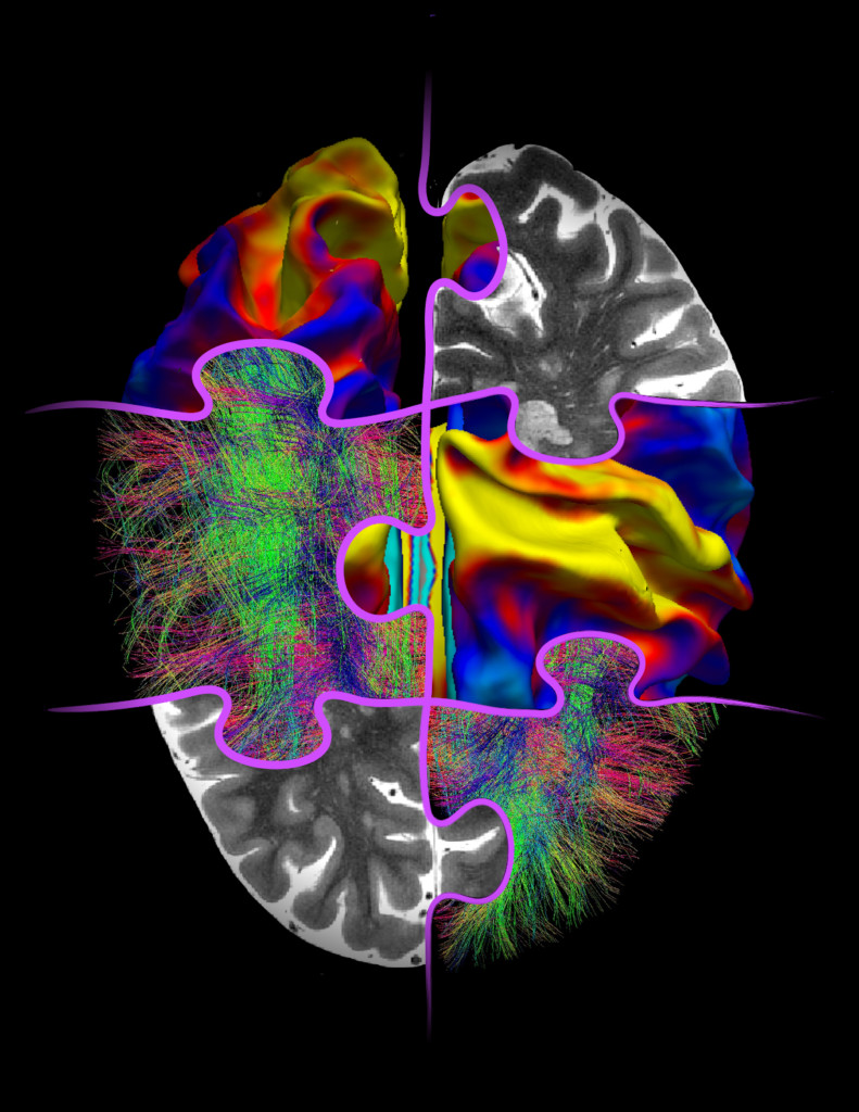
High-field magnetic resonance imaging (in black and white) distinguishes very small and subtle brain lesions. Surface-based imaging (blue-yellow-red) is used to easily study cortical tissue despite the cir convoluted shape of the brain. Finally, diffusion MRI (green-blue and red fibers) gives an estimate of the nerve fibers tracts that make up the white matter of the brain and connect to each other in the cortical ribbon.
In the case of multiple sclerosis, several neurodegenerative mechanisms take place at the same time. For example, myelin loss, diffuse inflammation or axon death. The interactions between these mechanisms are still poorly understood. Multimodal MRI makes it possible to study these mechanisms with great specificity and is therefore a technique of choice for solving the puzzle of the pathological process of multiple sclerosis.