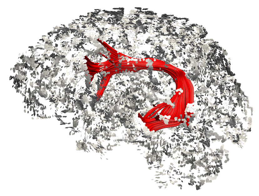
This picture originates from a severe Broca aphasia patient that regained the ability to speak after stroke. The image shows the underlying connectional anatomy of the language system obtained with MRI-based diffusion tensor imaging. The left hemisphere is shown as dots of the cortical layer. The image was created using the diffusion tensor imaging tractography software Trackvis (www.trackvis.org).